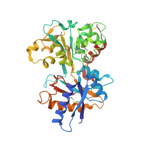
3FGSA

Crystal structure of g65r/k206e double mutant of the n-lobe human transferrin
Note, that the numbers in the matrix denote the consecutive residues in the loop, not the index of amino acids in the chain!
Knot K 2x +31
Knot core range Knot core length Knot tails range Slipknot tails range Slipknot loops range N-end length C-end length Type
+ 31 19-159 141 1-18, 160-220 18 60 slipknot
Fingerprint Knot forming loop Loop type
K +31 Asp63 <->
Bridging ionFe402
<-> Tyr188 ... Cys331 <-> Cys137 ... Asp63
ion-based
K +31 Asp63 <->
Bridging ionFe402
<-> Tyr188 ... Cys227 <-> Cys241 ... Cys331 <-> Cys137 ... Asp63
ion-based
Chain Sequence
DKTVRWCAVSEHEATKCQSFRDHMKSVIPSDGPSVACVKKASYLDCIRAIAANEADAVTLDARLVYDAYLAPNNLKPVVAEFYGSKEDPQTFYYAVAVVKKDSGFQMNQLRGKKSCHTGLGRSAGWNIPIGLLYCDLPEPRKPLEKAVANFFSGSCAPCADGTDFPQLCQLCPGCGCSTLNQYFGYSGAFKCLKDGAGDVAFVEHSTIFENLANKADRDQYELLCLDNTRKPVDEYKDCHLAQVPSHTVVARSMGGKEDLIWELLNQAQEHFGKDKSKEFQLFSSPHGKDLLFKDSAHGFLKVPPRMDAKMYLGYEYVTAIRNLREGTC
sequence length 329
structure length 329
publication title Structural and Functional Consequences of the Substitution of Glycine 65 with Arginine in the N-Lobe of Human Transferrin.
pubmed doi rcsb
molecule tags Metal transport
molecule keywords Serotransferrin
source organism Homo sapiens
total genus Genus: 99
ec nomenclature
pdb deposition date 2008-12-08
KnotProt deposition date 2018-10-31

pfam database annotations

chain Pfam Accession Code Pfam Family Identifier Pfam Description
A PF00405 Transferrin Transferrin
Image from the rcsb pdb (www.rcsb.org)
cath code
ClassArchitectureTopologyHomologyDomain
3.40.190.10 Alpha Beta 3-Layer(aba) Sandwich D-Maltodextrin-Binding Protein; domain 2 Periplasmic binding protein-like II 3fgsA01
3.40.190.10 Alpha Beta 3-Layer(aba) Sandwich D-Maltodextrin-Binding Protein; domain 2 Periplasmic binding protein-like II 3fgsA02
3.40.190.10 Alpha Beta 3-Layer(aba) Sandwich D-Maltodextrin-Binding Protein; domain 2 Periplasmic binding protein-like II 3fgsA01
3.40.190.10 Alpha Beta 3-Layer(aba) Sandwich D-Maltodextrin-Binding Protein; domain 2 Periplasmic binding protein-like II 3fgsA02
1FQFA 1N7WA 1D4NA 2O84X 3O97A 1A8EA 1FQEA 1B3EA 1N84A 1OQGA 1JQFA 1DTGA 2O7UA 1D3KA 1A8FA 3FGSA 1OQHA 1N7XA
chains in the KnotProt database with same CATH superfamily
1FQFA 1N7WA 1D4NA 2O84X 3O97A 1A8EA 1FQEA 1B3EA 1N84A 1OQGA 1JQFA 1DTGA 2O7UA 1D3KA 1A8FA 3FGSA 1OQHA 1N7XA
chains in the KnotProt database with same CATH topology
1FQFA 1N7WA 1D4NA 2O84X 3O97A 1A8EA 1FQEA 1B3EA 1N84A 1OQGA 1JQFA 1DTGA 2O7UA 1D3KA 1A8FA 3FGSA 1OQHA 1N7XA
chains in the KnotProt database with same CATH homology


#chains in the KnotProt database with same CATH superfamily
 1FQF A;  1N7W A;  1D4N A;  2O84 X;  3O97 A;  1A8E A;  1FQE A;  1B3E A;  1N84 A;  1OQG A;  1JQF A;  1DTG A;  2O7U A;  1D3K A;  1A8F A;  3FGS A;  1OQH A;  1N7X A; 
#chains in the KnotProt database with same CATH topology
 1FQF A;  1N7W A;  1D4N A;  2O84 X;  3O97 A;  1A8E A;  1FQE A;  1B3E A;  1N84 A;  1OQG A;  1JQF A;  1DTG A;  2O7U A;  1D3K A;  1A8F A;  3FGS A;  1OQH A;  1N7X A; 
#chains in the KnotProt database with same CATH homology
 1FQF A;  1N7W A;  1D4N A;  2O84 X;  3O97 A;  1A8E A;  1FQE A;  1B3E A;  1N84 A;  1OQG A;  1JQF A;  1DTG A;  2O7U A;  1D3K A;  1A8F A;  3FGS A;  1OQH A;  1N7X A; 
...loading similar chains, please wait...
similar chains in the KnotProt database (?% sequence similarity)
...loading similar chains, please wait...
similar chains in the pdb database (?% sequence similarity)

 
#similar chains in the KnotProt database (?% sequence similarity)
...loading similar chains, please wait...
#similar chains, but unknotted
...loading similar chains, please wait...
#similar chains in the pdb database (?% sequence similarity)
...loading similar chains, please wait...

KnotProt | Interdisciplinary Laboratory of Biological Systems Modelling